The latest updates issued by Philips assert the growing potential of its revolutionary lung suite in diagnosing and treating cancer at an early stage with the addition of global clinical partners.
Lung cancer is caused by the destruction of the lungs and lymph nodes when exposed directly or indirectly to hazardous substances such as smoke and chemicals.
Early symptoms of lung cancer, such as continuous coughing, muscle deficiency, discomfort in the chest, fatigue, losing appetite, and shortness of breath, are similar to cold symptoms and should be detected early to provide accurate therapy.
So, the lung suite launched by Philips with modern facilities makes it easier to identify and treat lung cancer at its initial stages.
Advanced features of Philips lung suite attract global partners
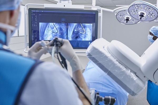
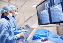
Addressing the need for early detection and treatment of lung cancer, Philips’ lung suite provides an all-in-one solution in a single room. The lung suite’s innovative real-time 3D imaging platform makes it easier for doctors to perform biopsies, resections, lesion marking, and Thoracic surgery in one room. This innovative approach by Philips has sparked a global interest in clinical entities resulting in the growth of networks for better care and standard facilities for patients. The latest update by Phillips lists numerous additions of partners in the revolutionary approach.
Several installations of Philips lung suite’s real-time 3D imaging solution were observed globally with a successful diagnosis of patients. Clinical entities from Belgium, France, Israel, and the UK have shown keen interest in the all-in-one suite by Philips. Some of the partners include the Royal Brompton Hospital in London, Hôpital Erasme in Brussels, Ziekenhuis Oost-Limburg (ZOL) Medical Center in Genk, and Carmel Medical Center in Haifa. Rouen University Hospital from Rouen, France, has also exhibited plans for using the innovative solution.
Embark of Philips’ lung suite in cancer treatment
With the advanced features Image Guided Therapy System, Philips’ lung suite embarks on providing minimally invasive therapy with precise diagnosis. The augmented fluoroscopic images provided by the therapy systems such as Azurion in the lung suite provide a real-time 3-D view of lesions.
The high resolution, real-time view of anatomical structures and lesions through Cone Beam CT imaging systems and X-ray detectors make it easier for the clinician to perform a guided biopsy using a catheter. In addition, the accurate biopsy result can be taken after the position of the catheter is confirmed using the same imaging system.
This innovative solution facilitates the ability to locate minor lesions and characterize them accordingly to proceed with therapy. The single-procedure therapy that includes the detection of lesions through bronchoscopy, ablation procedures, and image-guided surgery helps diagnose and treat lung cancer at the initial stage, improving the odds of survival. With the addition of revolutionary clinical partners, Philips aims to advance in providing standard medical care and a minimally interfering solution for patients with lung cancer.
Growing potential of lung suite for an accurate treatment of disease
The early-stage diagnosis of Cancer with Philips’ lung suite makes it possible to immediately treat patients according to the clinical procedures in one place. The advanced Imaging systems integrated with modern software offer precise Cone Beam CT imaging for nodules as small as 20mm. The lung suite detects peripheral lung nodules regardless of their positions and characteristics and allows clinicians to reach them safely for biopsy. Several successful first case trials signify the capability of this valuable tool in bronchoscopic microwave ablation of lesions.
The diagnostic potential of Philips lung suite indicated in the 2021 Journal of Bronchology & Interventional Pulmonology observes increased accuracy. This paper, written by researchers at Radboud University Medical Center, Netherlands, states an 18% rising accuracy of navigational bronchoscopy to reach target lesions, reducing more than half the total effective radiation doses per procedure.
Successful diagnosis and treatment of lung cancer patients were carried out at preliminary trials by Dr Kelvin Lau at St Bartholomew’s Hospital, London, using the lung suite. Additionally, several hospitals and established clinical facilities are expected to start the trials of this revolutionary solution.
An accurate diagnosis followed by successful treatment is essential in the early stages of lung cancer for the betterment of the patient. Unfortunately, non-small cell lung cancer (NSCLC) and small cell lung cancer (SCLC) become difficult to treat once the cells are damaged. Philips Lung suite makes it possible to precisely detect and treat initial stage lung cancer to improve the odds of survival and provide better facilities.


