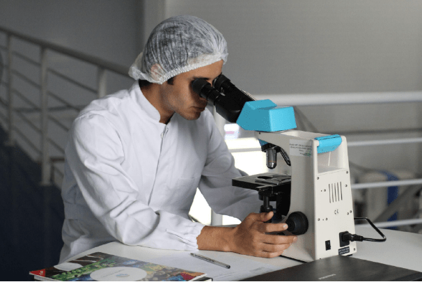
The global leader in scientific research, Thermo Fisher Scientific, installed the most futuristic cryo-transmission electron microscope in Hyderabad at the Centre for Cellular andMolecular Biology (CCMB) on April 12 th , 2022.
Thermo Fisher’s state-of-the-art cryo-electron microscopes are expected to boost extensive
study and help scientists step up their diagnostics research along with potential cures and
drug discoveries. Cryo-EM is an approach that ascertains the shape of a freezing sample to
preserve the natural structure of biological specimens by firing electrons at them and
recording the resulting images. Dr. Shekhar Mande, Director-General, Council of Scientific and Industrial Research (CSIR), kick-started the new facility. Researchers under CCMB and CSIR can utilize this facility together with researchers from other universities, institutes, pharma, and biotech companies.
What is a Cryo-Electron Microscope?
Transmission electron microscopes (TEM) use a beam of electrons to study the structure of
molecules and materials at the atomic scale. The wavelength of electrons being shorter than light can show minute details better than the super-resolution light microscopy.
However, few materials are not in accord with the high vacuum conditions and the intense
traditional beams used in TEMs. The water that surrounds the molecules evaporates, resulting in the burning of the high-power electrons, destroying them later on. To solve these problems, Cryo-EM uses frozen and gentler beams along with advanced image processing.
Thermo Fisher’s Scientific Cryo-EM samples can hold colder conditions for more than 72
hours inside the Krios Cryo-TEM. Within the Glacios or Tundra Cryo-TEM, the temperature
can be sustained for up to 24 hours. This allows scientists to gather the required data that can be used to generate a high-quality, effective reconstruction possible.
Working of Cryo-EM
Structural Biology is being replaced with Cryo-EM. It cools the specimens to colder
temperatures very quickly and thus preventing the water or H 2 O molecules from solidifying, preserving the original specimen structure. Once it freezes, numerous EM procedures can be applied to envision the sample in 3D in various resolutions, which includes the resolution of the architecture of the metabolic enzyme tied to the drug that disrupts its activity enabling researchers to get a deeper understanding.
This technique used by Thermo-Fisher’s Scientific Solutions comprises protein objects that
are frozen a few hours before exposing them to cryogenic temperatures or other biomolecules and then striking them with electrons to produce microscopic images of the specific molecules. They are used to redoing the 3D shape of a molecule which is useful for finding how proteins work and how they break down in diseases.
Advantages of Cryo-EM?
Using traditional methods like biophysics and Proton Magnetic resonance, also called NMR,
biologists who are interested in having a thorough knowledge of proteins as well as a
collection of proteins that interact with each other face numerous difficulties as proteins are
organically complex and tough to inspect. Those biologists who are curious to know more about proteins and the collection of proteins that interact with each other come across several challenges as proteins are organically complex and hard to inspect through traditional methods like biophysics and Nuclear Magnetic resonance (NMR).
Researchers can study proteins in their complex formations, structures, and changed forms. They can have a look at the several protein layouts in a single specimen. The resolution is improving, and the freezing method that is used is advanced. Hence, Cryo-EM is so fast and efficient that it can easily replace X-Ray crystallography in different ways.Cryo-EM does not need crystals and reduces radiation damage. Cryo-EM allows scientists to investigate the 3D protein structures and protein functions inside them which is important to understand how they operate, their roleplay in health hazards, and how they will react to therapies. It has become essential for scientists as it helps them create developments in research for communicable diseases, neurodegenerative diseases, and cancer.
Thermo Fisher’s Role in Cryo-EM Research and Discoveries
The setup of the new facility by Thermo Fisher Solutions offers a suite of mechanical and
specimen handling tech, and this increases easy usage and guarantees the ultimate amount of superior data that can be collected for every single sample. Amit Chopra, managing director, India, and South Asia, Thermo-Fisher Scientific, stated that the induction of Cryo-EM microscopes by them will benefit CCMB to scrutinize macromolecular structures and create a research awareness foundation and skills for Cryo-EM study in India. The study of human biology, enzymology, and drug discovery will be
encouraged through this facility. The technology at the new facility will help researchers to function with models at cryogenic temperatures, (around -173°C), and image-specific molecules. Also, apart from this, the addition of Cryo-EM microscopy makes it an exceptional facility for researchers to investigate interesting details of living cells that were never done before.


